Diagram Of Bones In Neck And Shoulder / Bones Of The Skull And Shoulder Girdle Ppt Video Online Download - The neurocranium is the part enveloping the brain and is formed out of two parts;
byAdmin-
0
Diagram Of Bones In Neck And Shoulder / Bones Of The Skull And Shoulder Girdle Ppt Video Online Download - The neurocranium is the part enveloping the brain and is formed out of two parts;. The neurocranium (cranial vault) and the viscerocranium (facial skeleton). When this condition develops in the cervical spine, it is called cervical osteoarthritis. Shoulder joint of human body anatomy infographic diagram with all parts including bones ligaments muscles bursa cavity capsule cartilage membrane for medical science. Contains glands ( thyroid, parathyroid, and thymus ), the larynx, pharynx and trachea. In this video part, you will also find out the anatomy of the neck and shoulders.
Contains glands ( thyroid, parathyroid, and thymus ), the larynx, pharynx and trachea. The shoulder is a complex combination of bones and joints where many muscles act to provide the widest range of motion of any part of the body. The column of the neck bones is slightly curved. Shoulder joint of human body anatomy infographic diagram with all parts including bones ligaments muscles bursa cavity capsule cartilage membrane for medical science. Most superficial muscle covering posterior shoulder innervation:
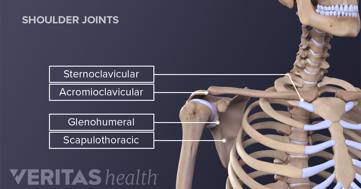
Neck Anatomy Pictures Bones Muscles Nerves from www.healthpages.org Axial skeleton — bones of the skull, vertebral column, thoracic cage. These critical parts of the upper body are very prone to developing pain because the position of all the bones in the neck and shoulders are completely dependent on the balance and alignment of the muscles and fascia that lash them together and allow for movement between them. The most common descriptions for collarbone pain are tender, throbbing, aching, dull, or stabbing. A second joint in the shoulder is the junction of the collar bone with the shoulder blade, called. Most superficial muscle covering posterior shoulder innervation: Accessory nerve spinal part (cn xi) nerve runs underneath entire length of muscle beginning at the bast of skull and posterolateral surface of the neck can be pinched by a blow to the neck in martial arts (stuns the nerve) The first one that holds the skull is called the atlas. The column of the neck bones is slightly curved.
Cervical spine anatomy is quite complex.
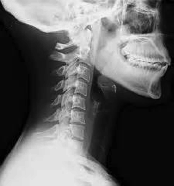
Cervical radiculopathy, commonly called a pinched nerve occurs when a nerve in the neck is compressed or irritated where it branches away from the spinal cord. The neck is unique in that it supports the weight of your head (10 to 11 pounds) and allows a variety of head/neck movement, such as turning your head from side to. Cervical osteoarthritis (neck arthritis) osteoarthritis occurs when the protective cartilage in a joint begins to break down and no longer facilitates smooth movement between bones, which can eventually result in the joint becoming swollen and painful. Diagram of bones in neck and shoulder. Innerbody research is the largest home health and wellness guide online, helping over one million visitors each month learn about health products and services. Bones of the neck picture. Shoulder joint of human body anatomy infographic diagram with all parts including bones ligaments muscles bursa cavity capsule cartilage membrane for medical science. The neck bones are called the cervical vertebrae. There are seven of them. In this video part, you will also find out the anatomy of the neck and shoulders. There are seven cervical vertebrae that allow for a great amount of motion in the neck. Neck and shoulder pain anatomy. We will attempt to provide a simplified overview of this complex anatomy.
The column of the neck bones is slightly curved. There are seven of them. Innerbody research is the largest home health and wellness guide online, helping over one million visitors each month learn about health products and services. Each arm is attached to a shoulder blade. We will attempt to provide a simplified overview of this complex anatomy.
Shoulder Joint Structure from embed.widencdn.net Shoulder girdle , radiographs : Bones of the neck picture. There are seven cervical vertebrae that allow for a great amount of motion in the neck. Diagram of bones in neck and shoulder. It consists of two major parts: The shoulder is a complex combination of bones and joints where many muscles act to provide the widest range of motion of any part of the body. Cervical osteoarthritis (neck arthritis) osteoarthritis occurs when the protective cartilage in a joint begins to break down and no longer facilitates smooth movement between bones, which can eventually result in the joint becoming swollen and painful. The first one that holds the skull is called the atlas.
The skull base that supports the brain and the calvaria (skullcap) that sits on top of the base, covering the brain.
When this condition develops in the cervical spine, it is called cervical osteoarthritis. The neck and shoulders are complex and interconnected areas, and medical problems that affect one often affect the other, as well. Axial skeleton — bones of the skull, vertebral column, thoracic cage. Bones of the head and neck. These bones have some interesting landmarks, including various bumps and projections. The bones of the shoulder consist of the humerus (the upper arm bone), the scapula (the shoulder it forms. Shoulder anatomy sketch, scapula and humerus bone human shoulder anatomy with vector sketch of scapula and humerus bones, medicine and health care design. Contains cervical vertebrae and postural muscles. Innerbody research is the largest home health and wellness guide online, helping over one million visitors each month learn about health products and services. The skull is a strong, bony capsule that rests on the neck and encloses the brain. The first one that holds the skull is called the atlas. Most superficial muscle covering posterior shoulder innervation: The skull base that supports the brain and the calvaria (skullcap) that sits on top of the base, covering the brain.
The most common causes of collarbone pain are related to injuries. Although anchored in the neck, their primary functions are to move the shoulder blades and support the arms. However, collarbone pain can come on with or without injury, gradually or suddenly. Boneka stitch jumbo warna biru. The most common descriptions for collarbone pain are tender, throbbing, aching, dull, or stabbing.
Bones Of The Upper Limb Ul And The Three Joints Of The Shoulder Download Scientific Diagram from www.researchgate.net Shoulder girdle an overview sciencedirect topics. The neurocranium (cranial vault) and the viscerocranium (facial skeleton). In this video part, you will also find out the anatomy of the neck and shoulders. The bones of the head and neck play the vital role of supporting the brain, sensory organs, nerves, and blood vessels of the head and protecting these structures from mechanical damage. The neck is unique in that it supports the weight of your head (10 to 11 pounds) and allows a variety of head/neck movement, such as turning your head from side to. Boneka stitch jumbo warna biru. Most superficial muscle covering posterior shoulder innervation: There are seven cervical vertebrae that allow for a great amount of motion in the neck.
However, collarbone pain can come on with or without injury, gradually or suddenly.
The neck bones are called the cervical vertebrae. Cervical spine anatomy is quite complex. In adults the long bones of the legs and arms are filled with yellow marrow. #bones in the head neck and shoulder #bones of the head neck and shoulder girdle #bones of the head neck and shoulders #function of the head neck and shoulder bones #the position of the head face neck chest and shoulder girdle bones This may cause pain that radiates into the shoulder, as well as numbness that travels down the arm and into the hand. The most common causes of collarbone pain are related to injuries. Bones of the head and neck. Axial skeleton — bones of the skull, vertebral column, thoracic cage. Shoulder joint of human body anatomy infographic diagram with all parts including bones ligaments muscles bursa cavity capsule cartilage membrane for medical science. The skull is a strong, bony capsule that rests on the neck and encloses the brain. This will give depth to the mouth and allow the portrait to seem more natural. A second joint in the shoulder is the junction of the collar bone with the shoulder blade, called. There are seven of them.